Radiology Services at Greater Lancashire Hospital
Expert MRI Scans…
At Greater Lancashire Hospital, we offer advanced MRI scans to diagnose conditions, plan treatments, and assess treatment effectiveness. Using powerful magnets and radio waves, MRI scans provide detailed images of the brain, spinal cord, bones, joints, and internal organs like the liver and heart. The procedure is safe, non-invasive, and performed by trained radiographers who guide you through each step. MRI results help ensure accurate diagnoses and effective treatment planning for a range of medical conditions.
What is an MRI scan?
Magnetic resonance imaging (MRI) is a type of scan that uses strong magnetic fields and radio waves to produce detailed images of the inside of the body.
An MRI scanner is a large tube that contains powerful magnets. You lie inside the tube during the scan.
The results of an MRI scan can be used to help diagnose conditions, plan treatments and assess how effective previous treatment has been.
MRI Scans can be used to scan the:
- Brain
- Spinal cord
- Bones and joints
- Breasts
- Heart
- Blood vessels
- Internal organs, such as the liver, womb or prostate gland
What happens during an MRI Scan
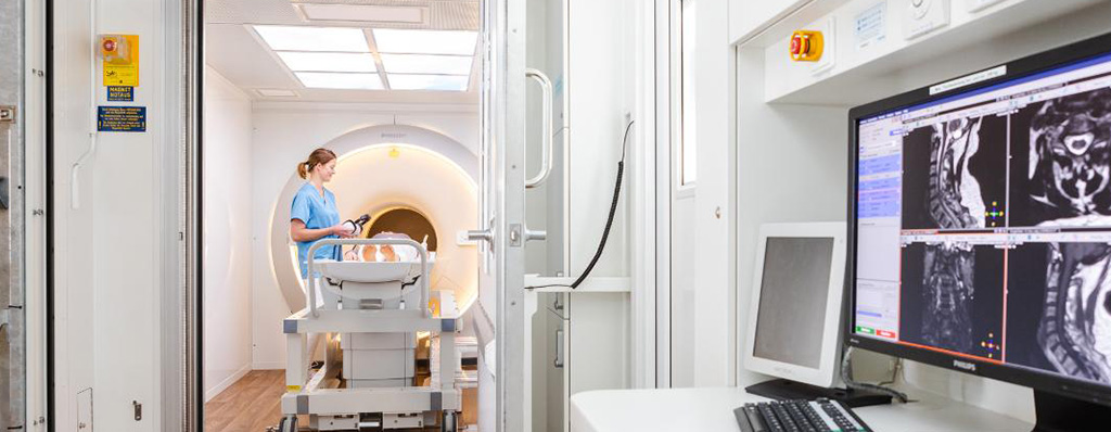
During an MRI scan, you lie on a flat bed that’s moved into the scanner.
Depending on the part of your body being scanned, you’ll be moved into the scanner either head first or feet first.
The MRI scanner is operated by a radiographer, who is trained in carrying out imaging investigations.
They control the scanner using a computer, which is in a different room, to keep it away from the magnetic field generated by the scanner.
You’ll be able to talk to the radiographer through an intercom and they’ll be able to see you on a television monitor and through the viewing window throughout the scan.
At certain times during the scan, the scanner will make loud tapping noises. This is the electric current in the scanner coils being turned on and off.
You’ll be given earplugs or headphones to wear.
It’s very important to keep as still as possible during your MRI scan.
The radiographer may ask you to hold your breath for a few seconds or follow other instructions during the scan. The scan lasts 15 to 90 minutes, depending on the size of the area being scanned and how many images are taken.
How does an MRI scan work?
Most of the human body is made up of water molecules, which consist of hydrogen and oxygen atoms.
At the centre of each hydrogen atom is an even smaller particle called a proton. Protons are like tiny magnets and are very sensitive to magnetic fields.
When you lie under the powerful scanner magnets, the protons in your body line up in the same direction, in the same way that a magnet can pull the needle of a compass. You will not be able to feel this.
Short bursts of radio waves are then sent to certain areas of the body, knocking the protons out of alignment.
When the radio waves are turned off, the protons realign. This sends out radio signals, which are picked up by receivers.
These signals provide information about the exact location of the protons in the body.
They also help to distinguish between the various types of tissue in the body, because the protons in different types of tissue realign at different speeds and produce distinct signals.
In the same way that millions of pixels on a computer screen can create complex pictures, the signals from the millions of protons in the body are combined to create a detailed image of the inside of the body.
Expert Ultrasound Scans…
We also provide safe and effective ultrasound scans to diagnose conditions, monitor pregnancies, and assist in surgical procedures. Using high-frequency sound waves, ultrasound creates real-time images of the inside of the body. The procedure is non-invasive, with an ultrasound probe emitting sound waves that bounce back, creating an image displayed on a monitor. Ultrasound scans can be used to examine various areas such as the heart, organs, muscles, and joints. Whether external or internal, our trained professionals ensure a comfortable experience for all patients.
What is an Ultrasound Scan & how do they work?
An ultrasound scan, sometimes called a sonogram, is a procedure that uses high-frequency sound waves to create an image of part of the inside of the body.
An ultrasound scan can be used to monitor an unborn baby, diagnose a condition, or guide a surgeon during certain procedures.
A small device called an ultrasound probe is used, which gives off high-frequency sound waves.
You can’t hear these sound waves, but when they bounce off different parts of the body, they create “echoes” that are picked up by the probe and turned into a moving image. This image is displayed on a monitor while the scan is carried out.
Ultrasound types & uses
External ultrasound scan
An external ultrasound scan is most often used to examine the heart, organs in the tummy and pelvis, muscles and joints and other organs and tissues that can assessed through the skin.
Internal ultrasound scan
An internal examination allows a doctor to look more closely inside the body at organs such as the prostate gland, ovaries or womb.
Preparing for an ultrasound scan
Before having some types of ultrasound scan, you may be asked to follow certain instructions to help improve the quality of the images produced.
For example, you may be advised to:
- drink water and not go to the toilet until after the scan – this may be needed before a scan of your unborn baby or your pelvic area
- avoid eating or drinking for several hours before the scan – this may be needed before a scan of your digestive system, including the liver and gallbladder
Depending on the area of your body being examined, the hospital may ask you to remove some clothing and wear a hospital gown.
What happens during an Ultrasound?
Most ultrasound scans last between 15 and 45 minutes. They usually take place in a hospital radiology department and are performed either by a doctor (radiologist) or a sonographer.
They can also be carried out in community locations such as GP practices, and may be performed by other healthcare professionals, such as midwives or physiotherapists who have been specially trained in ultrasound.
There are different kinds of ultrasound scans, depending on which part of the body is being scanned and why.
The 3 main types are:
- Endoscopic ultrasound scan – the probe is attached to a long, thin, flexible tube (an endoscope) and passed further into the body – This type of scan is not undertaken at Greater Lancashire Hospital
- External ultrasound scan – the probe is moved over the skin
- Internal ultrasound scan – the probe is inserted into the body.